Functional Near-Infrared Spectroscopy (fNIRS)
Functional Near-Infrared Spectroscopy Defined
(fNIRS)
What Is Functional Near-Infrared Spectroscopy?
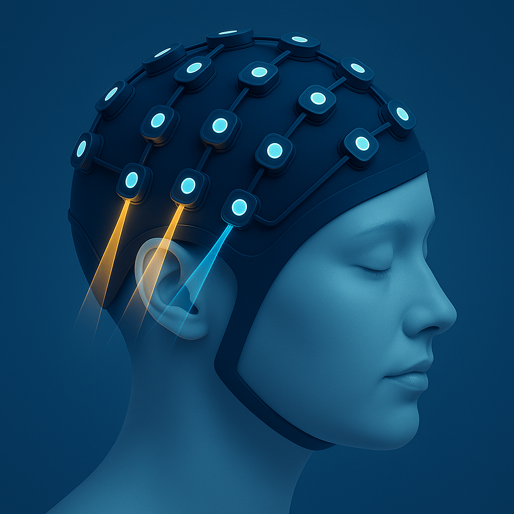
fNIRS is a non-invasive optical imaging technique that uses near-infrared light to measure changes in blood oxygen levels in the brain. It provides insights into brain activity by detecting the hemodynamic responses associated with neural activation.
What Does fNIRS Treat?
fNIRS is not used for treatment, but rather in cognitive neuroscience, psychology, and rehabilitation studies by mapping brain function and monitoring neural responses in real-time. It is commonly used for:
Cognitive and Neural Research: Assessing brain function in both healthy individuals and those with neurological or psychiatric disorders.
Brain-Computer Interfaces (BCIs): Exploring communication methods for individuals with severe disabilities.
Pediatric Neurology: Studying developmental disorders like autism and ADHD.
fNIRS Mechanism of Action
Near-infrared light is emitted through the scalp, penetrating brain tissue. Detectors measure the light that is reflected back, which varies as the concentrations of oxy-hemoglobin (HbO) and de-oxy hemoglobin (HbR) change with brain activity. Algorithms then process this data to map how brain activity responds to tasks or stimuli.
Is fNIRS FDA Approved?
Some fNIRS devices are FDA-cleared for diagnostic and monitoring purposes, but it is not FDA-approved as a treatment.
Can fNIRS Treat Alzheimer’s?
Though not used as a treatment for Alzheimer’s, fNIRS can be used alongside stimulation methods to observe their affect on activity in the target brain region.
It is not therapeutic, but it may guide future interventions, including TEMT-RF.
-
fNIRS does not “target” brain regions the way stimulation methods do, because it is a method of observing rather than affecting the brain. Because infrared light can only penetrate a few centimeters into tissue, fNIRS focuses on superficial regions such as the prefrontal, motor, and somatosensory cortexes.
-
As a research tool, fNIRS it is used to study the disease by:
Monitoring Brain Activity: Identifying regions with altered hemodynamics in individuals with Alzheimer’s disease.
Assessing Cognitive Decline: Evaluating changes in neural activity during memory and problem-solving tasks.
Early Detection: Investigating biomarkers for earlier diagnosis of Alzheimer’s and related dementias.
-
Recent studies have explored using fNIRS to detect preclinical stages of Alzheimer’s by observing hemodynamic changes during cognitive tasks.
How does fNIRS compare to TEMT-RF?
Functional Near-Infrared Spectroscopy (fNIRS) and Transcranial Electromagnetic Treatment with Radio Frequencies (TEMT-RF) are fundamentally different technologies with distinct purposes.
TEMT-RF uses ultra-high frequency radio waves to modulate brain function and cellular activity delivered through a non-invasive wearable headset.
Functional Near-Infrared Spectroscopy (fNIRS) is a brain imaging technique that measures changes in blood oxygenation and hemodynamics to infer neural activity through a wearable, potentially portable headset.
Learn more about Functional Near-Infrared Spectroscopy
Functional Near-Infrared Spectroscopy Products
Functional Near-Infrared Spectroscopy Research
Alzheimer’s Research
Early Detection of Alzheimer’s Disease Using Non-invasive Near-Infrared Spectroscopy
Functional near-infrared spectroscopy in elderly patients with four types of dementia
Other Research
Investigating Mild Cognitive Impairment in Patients And Controls With TD-fNIRS
A Study to Asses Wellness Using a Brain Sensing Device on Physicians
Assessing the Effects of the Muse Meditation System on Cognition and Well-being (MuseCog)
Meditation Utilizing Signals From Electroencephalography in Chronic Pain (MUSE-PAIN) Study
Cortical Signal Analysis and Advances in Functional Near-Infrared Spectroscopy Signal: A Review
External links are provided for reference only and do not imply affiliation, endorsement, or recommendation by NeuroEM Therapeutics. NeuroEM is not responsible for the content, accuracy, or claims made on external sites. All trademarks and copyrights are the property of their respective owners.